Advanced Diagnostic Imaging
Echocardiography (Cardiac Ultrasound)
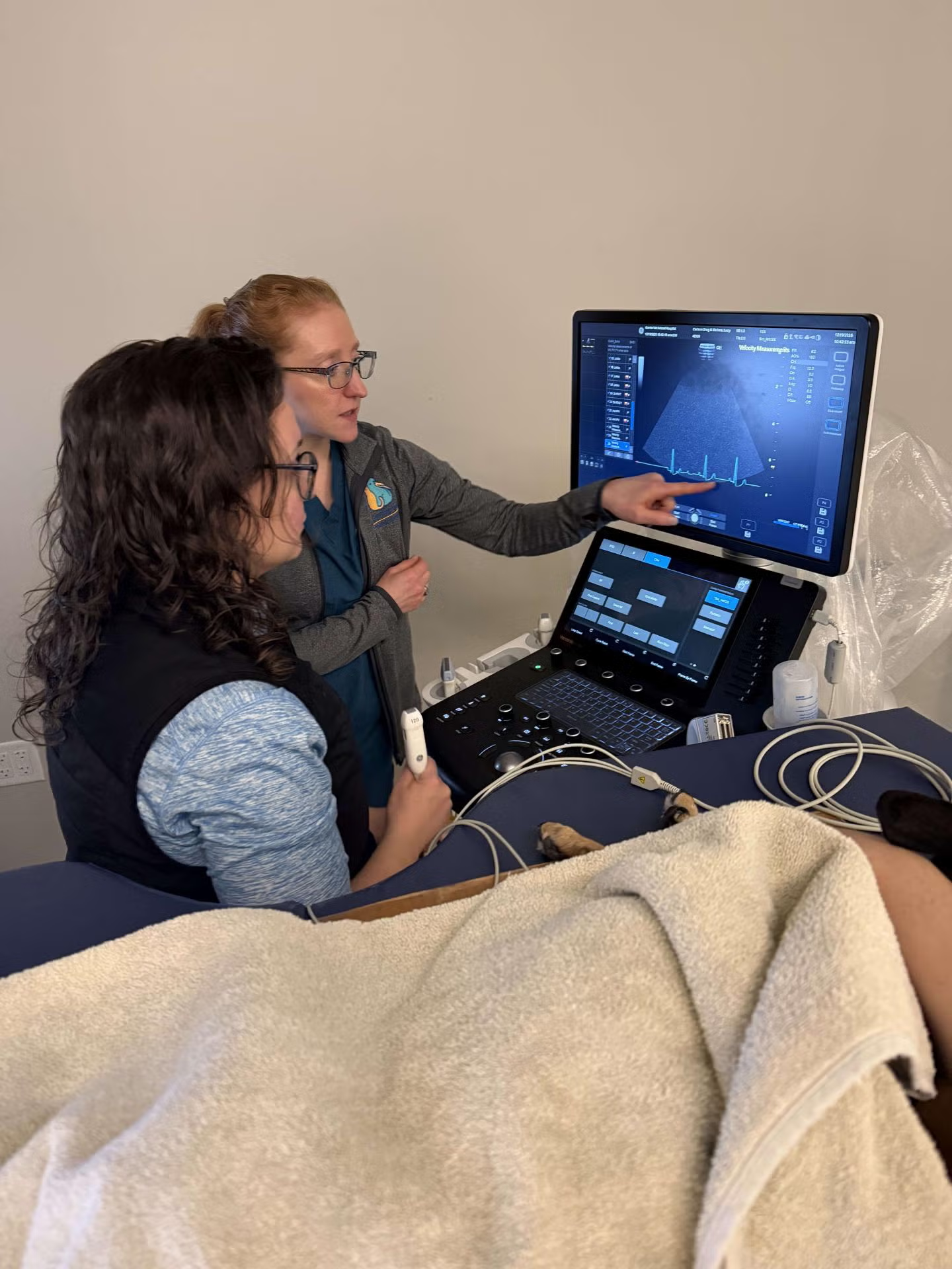
Echocardiography is one of the most valuable diagnostic tools used in veterinary medicine to evaluate heart structure and function in animals. Using ultrasound waves, we can visualize the heart in real time to assess chamber size, wall thickness, valve structure, blood flow, and overall cardiac performance.
This non-invasive imaging technique helps diagnose conditions such as dilated cardiomyopathy, hypertrophic cardiomyopathy, congenital heart defects, and valvular disease.
Our staff has completed specialized training in veterinary cardiac ultrasound, allowing them to obtain high-quality diagnostic images and perform detailed cardiac measurements that are essential for accurate evaluation. An additional benefit is that these studies can be reviewed and interpreted by a board-certified veterinary cardiologist, providing an expert assessment and helping ensure the most accurate diagnosis and treatment recommendations for each patient.
Early Detection for At-Risk Breeds
Cardiac ultrasound is also an important tool for early screening in breeds known to be at higher risk for inherited heart disease. Breeds such as Doberman Pinschers, Boxers, Maine Coon cats, and Cavalier King Charles Spaniels may develop cardiac disease that begins with very subtle changes long before clinical signs appear.
Routine echocardiographic screening can detect early abnormalities—such as mild chamber enlargement, reduced contractility, or early valve changes—allowing veterinarians to monitor patients closely and intervene sooner when needed. Early detection can help guide lifestyle and medical management decisions that may slow disease progression and support a longer, better quality of life for affected pets.
Ultrasonography (Diagnostic Ultrasound)
Ultrasonography is an advanced diagnostic imaging technique that uses high-frequency sound waves to create real-time images of structures inside your pet's body.
Unlike some other imaging methods, such as X-rays, ultrasound does not use radiation. Instead, a specialized probe sends sound waves into the body, and the returning echoes are used to generate detailed images of organs and tissues. This allows veterinarians to evaluate structures such as the heart, liver, kidneys, spleen, bladder, and other soft tissues in a completely non-invasive and painless way.
Our team has completed specialized training in veterinary ultrasonography to ensure that studies are performed with precision and high diagnostic quality. When indicated, ultrasound images may also be reviewed by a board-certified radiologist to provide an additional expert interpretation.
Ultrasound-Guided Procedures
In addition to diagnosing many common diseases, ultrasound can also be used to guide minimally invasive procedures such as fine-needle aspirates or tissue biopsies. By using ultrasound guidance, veterinarians can precisely target abnormal areas while avoiding surrounding structures, often allowing diagnostic samples to be collected with less discomfort and lower risk compared to traditional surgical biopsy methods.
This collaborative, technology-driven approach ensures your pet receives accurate diagnosis, timely intervention, and the highest standard of compassionate cardiac care.